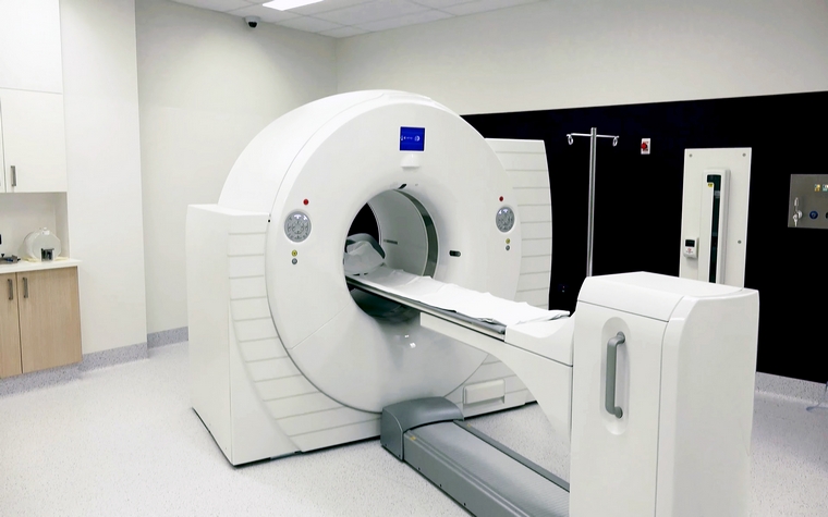
PET-MR is an imaging method that combines Positron Emission Tomography (PET) and Magnetic Resonance Imaging (MR) technologies. This hybrid technology combines the advantages of both methods to obtain more detailed and comprehensive images. PET-MR is used in the diagnosis and treatment monitoring of cancer, neurological, and cardiovascular diseases.
What is PET-MR?
PET-MR devices are hybrid imaging systems that utilize cutting-edge technology. These systems combine PET and MR imaging within a single machine. MR imaging, a crucial imaging method in radiology, functions like a giant magnet generating a magnetic field. Compared to CT technology, MRI has significant advantages such as not containing ionizing radiation, having a high ability to image soft tissue, and being able to obtain images in multiple planes. Combining these superior features with metabolic information (information related to the body's active functions) from PET has made PET-MR imaging prominent, particularly in oncology patients, for diagnosis, patient monitoring, and treatment evaluation. PET-MR combines metabolic information obtained from PET with anatomical and functional information from MR to provide a combined image. This image allows the specialist physician to obtain important information for the benefit of the patient. PET-MR devices are frequently used, especially for oncology patients. In oncology patients, PET-MR imaging serves three main purposes:
What is PET-MR used for?
- Used for identifying potential cancer foci and diagnosing cancer: This step involves scanning almost the entire body from head to toe. During this scan, MRI provides anatomical information (related to the structure of the organ being imaged) about the soft tissues. This information is combined with metabolic information obtained simultaneously through PET imaging. The result is a PET-MRI image. Since both pieces of information are obtained simultaneously and evaluated on a single image, if cancer is present, its focus can be identified with high accuracy. In this way, whole-body cancer screening can be performed as part of a check-up, thus enabling early diagnosis of potential cancer.
- It is used in the staging of detected cancer: Since the entire body can be scanned in a single session, distant metastasis staging can also be done simultaneously with the cancer diagnosis.
- It is used to evaluate the response to treatment in patients undergoing oncological treatment: The treated area containing the cancer is examined by considering both the anatomical information provided by high-powered MRI and the metabolic information provided by PET. This allows for the evaluation of both the size change resulting from treatment and the metabolic response to treatment. Since all of this can be done in a single session, patients are spared from having to undergo multiple different imaging methods. This also prevents time loss, which is a significant factor in oncological treatments.
Which diseases can be diagnosed using PET-MRI?
PET-MR plays an important role in the diagnosis of various diseases. Below is a list of the main diseases and conditions for which PET-MR is useful in diagnosis:
- Brain Tumors: PET-MR is highly effective in determining the location, size, and extent of spread of brain tumors. By showing both anatomical details and metabolic activity, it allows for a more accurate assessment of tumors.
- Lymphoma: PET-MR is used in the diagnosis and staging of lymphoma. It provides detailed imaging of lymph nodes and other tissues, which makes it possible to better understand the spread of the disease.
- Prostate Cancer: PET-MR is an important tool in the diagnosis and determination of the spread of prostate cancer. The detailed anatomical imaging capabilities of MR, combined with PET's ability to show metabolic activity, allow for more precise detection of cancerous tissue.
- Alzheimer's Disease: PET-MR is used in the early diagnosis of Alzheimer's disease. It helps monitor the progression of the disease by showing biochemical changes and anatomical structures in the brain.
- Parkinson's Disease: PET-MR is used in the diagnosis and staging of Parkinson's disease. It supports the diagnosis by evaluating dopamine activity and structural changes in the brain.
- Cardiovascular Diseases: PET-MR is effective in assessing the viability of the heart muscle and blood flow. It is used to identify damaged tissues after a heart attack and blockages in the arteries.
- Infections and Inflammatory Diseases: PET-MR can be used to detect infection foci and inflammatory processes in the body. This is particularly useful in complex cases and chronic inflammatory diseases.
Beyond these diseases, PET-MR can also be used to evaluate many other medical conditions and optimize treatment plans. This technology provides doctors with more comprehensive and accurate information about patients' conditions, making it a vital tool in the early diagnosis and treatment of diseases.
Is PET-MRI Safe?
Yes, PET-MR is generally a safe procedure. The MR portion uses radio waves and magnetic fields for imaging and therefore involves no radiation. The PET portion uses a low dose of radioactive material, but this dose is generally considered safe for patients. Nevertheless, careful consideration should be given to pregnant women and patients with certain special conditions.
Is PET-MR different from PET-CT?
Yes, PET-MR and PET-CT are different technologies. PET-CT is a combination of PET and Computed Tomography (CT). CT uses X-rays to image and show anatomical details. PET-MR, on the other hand, uses MRI to provide more detailed soft tissue images and reduces radiation exposure. Therefore, PET-MR may be preferred in some cases.
How long does a PET-MRI scan take?
A PET-MR scan typically takes 1 to 2 hours. The duration may vary depending on the scope of the examination and the patient's condition.
Is PET-MRI a painful procedure?
No, the PET-MR procedure is painless. However, some patients may experience slight discomfort because you need to remain still during the procedure. Also, a needle-pinching sensation may be felt when a radioactive substance is injected intravenously for the PET scan.
When will the PET-MR results be available?
PET-MR results are usually ready within a few days. The radiologist analyzes the images and reports the results. The results are communicated to the patient's doctor, who then informs the patient.
In which situations is PET-MR preferred over PET-CT?
PET-MR is preferred over PET-CT, especially in cases where soft tissue details are important. It provides superior anatomical detail in imaging organs such as the brain, prostate, and liver. Additionally, PET-MR may be more frequently preferred in children and young patients to reduce radiation exposure.
Is any preparation required before a PET-MRI?
Yes, some preparations may be necessary before a PET-MRI. For example, you may be asked to fast for a few hours before the procedure. It's also important to inform your doctor if you are taking any specific medications. Detailed preparation instructions will be provided to you before your appointment.
Is there radiation exposure during a PET-MRI?
Yes, you will be exposed to a low level of radiation during PET-MR because a radioactive substance is used. However, this dose is generally considered safe, and the MR portion itself is radiation-free. Nevertheless, necessary precautions are taken to minimize radiation exposure.


 Make an Appointment
 E-Result
 Let Us Call You