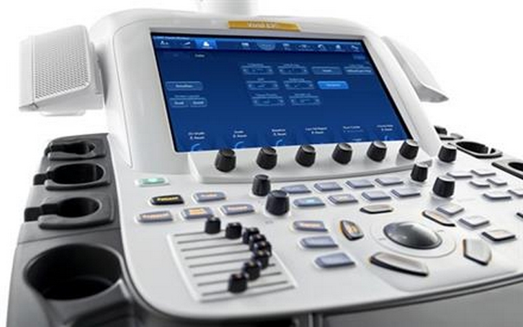
What is an Echocardiography (Echo) Device and What Does It Do?
An echocardiography device is a medical device used to image the heart and surrounding blood vessels. This device, which works with ultrasound technology, can examine the structure and functions of the heart in detail with the help of sound waves. Echocardiography helps cardiologists diagnose heart diseases and plan treatment processes.
What is Echocardiography?
Echocardiography is the process of imaging the heart and great vessels with ultrasound. This process is used to detect structural and functional disorders of the heart. Echocardiography is a non-invasive method and does not cause any harm to patients. For this reason, it is often preferred in the diagnosis of heart diseases.
What are the Echocardiography Methods?
Echocardiography has various methods used to evaluate the structure and function of the heart and vessels.
1. Two-Dimensional (2D) and Three-Dimensional (3D) Echocardiography
2D Echocardiography: Provides two-dimensional images of the heart walls, valves, and great vessels connected to the heart. Standard echocardiography usually begins with 2D imaging and is used to evaluate the overall structure and function of the heart.
3D Echocardiography: Available in some medical centers and hospitals, this method is used to obtain more detailed information about certain areas, especially the lower left chamber of the heart. The lower left chamber is the main pumping area of the heart, and 3D imaging allows for detailed examination of this area.
2. Doppler Echocardiography
Doppler echocardiography is based on the principle that sound waves reflect off blood cells within the heart and blood vessels, changing their pitch. These changes are called Doppler signals and measure the speed and direction of blood flow. Doppler echocardiography shows blockages or leaks in the heart valves and checks blood pressure in the heart arteries.
3. Color Flow Display
Color flow imaging shows blood flow through the heart in color. This helps identify leaky heart valves and other changes in blood flow. It provides a detailed analysis of the direction and speed of blood flow through the heart by showing it in color.
These echocardiography methods provide a detailed examination of cardiovascular health and provide cardiologists with important data to create accurate diagnoses and treatment plans.
How is an Echocardiogram Performed?
Echocardiography is performed in a supine position with the patient. Gel is applied to the skin to facilitate the movement of the ultrasound probe and increase the clarity of the images. The echocardiography probe is placed at different points on the chest to take images of the heart and blood vessels from different angles. The patient does not feel any pain during the procedure and the procedure usually takes 30-60 minutes.
What can be examined with an echo device?
The echo machine can examine the heart's structural abnormalities, valve function, heart wall movement, and blood flow within the heart. It can also assess the size of the heart chambers and the thickness of the heart muscle. This information provides cardiologists with important data to help them assess patients' heart health and develop treatment plans.
In Which Diseases Is Echocardiography Used?
Echocardiography is used to diagnose many heart diseases such as heart failure, heart valve diseases, congenital heart diseases, heart rhythm disorders and myocardial infarction. It also plays an important role in the evaluation of hypertension and other vascular diseases.
Does an Echocardiography Device Emits Radiation?
The echocardiography device does not emit radiation because it works with ultrasound technology. Therefore, echocardiography is a safe imaging method for patients. Since it does not carry a radiation risk, it can be used safely in children, pregnant women and other patient groups that are sensitive to radiation.
Does the Echocardiography Device Make Noise?
The echocardiography device is generally quiet and does not produce any noticeable noise that can be heard by the patient. A slight noise may be heard during movement of the ultrasound probe, but this noise is not at a level that will disturb the patient.


