What is the Radiology Department and What Diseases Does It Treat?
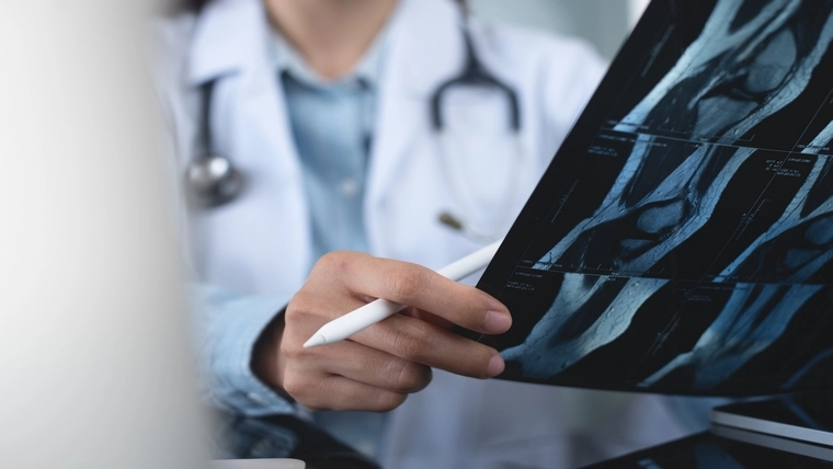
The radiology department is a branch of medicine that plays a critical role in the diagnosis and treatment of diseases using medical imaging techniques. In this department, organs and tissues within the body are imaged with methods such as X-ray, Magnetic Resonance Imaging (MRI), computed tomography (CT), Ultrasound, PET-MRI. Radiology is used in the diagnosis of a wide range of diseases, from cancer to bone fractures, from heart diseases to brain tumors.
About the Radiology Department
The radiology department has become an integral part of modern medicine and is indispensable for patients to receive accurate diagnosis and to be directed to effective treatment. Radiologists diagnose diseases using imaging methods and collaborate with other specialist doctors in light of this information. The department plays a key role in both the diagnosis and treatment process.
Who is a Radiology Specialist and What Are Their Duties?
A radiologist is a doctor who specializes in diagnosing and treating diseases using medical imaging techniques. Duties include performing imaging tests, analyzing images, and advising other doctors on diagnosis and treatment planning. Radiologists may also perform some therapeutic procedures called interventional radiology.
What Diseases Does Radiology Treat?
Radiology is a branch of medicine used to diagnose and treat diseases and looks at a wide range of diseases. Here are the main diseases that radiology plays a role in diagnosing and treating:
Cancers:- Lung Cancer: Radiology is used in the diagnosis of lung cancer, especially with X-ray and computerized tomography (CT) methods.
- Breast Cancer: Mammography and Magnetic Resonance Imaging (MRI) play a critical role in the early diagnosis of breast cancer.
- Brain Tumors: MRI and CT are commonly used to detect and monitor brain tumors.
- Prostate Cancer: Prostate MRI is used in the detection and treatment planning of prostate cancer.
- Coronary Artery Disease: Coronary CT angiography provides information about the condition of the heart vessels and is used to assess the risk of heart attack.
- Aortic Aneurysm: US and CT are used to evaluate aortic artery dilatations and the risk of rupture.
- Deep Vein Thrombosis: US is a common imaging method used to detect clots in the leg veins.
- Stroke: CT and MRI are used to determine the extent and location of damage in the brain after a stroke.
- Multiple Sclerosis (MS): MRI is used to detect lesions in the brain and spinal cord.
- Aneurysms and AVMs (Arteriovenous Malformations): MR angiography and CT are used to detect vascular abnormalities in the brain.
- Fractures and Cracks: X-ray is the first method used to detect bone fractures.
- Osteoarthritis: MRI and X-rays are used to determine the degree of joint degeneration and damage to the joint.
- Ligament and Tendon Injuries: MRI is an effective method for detecting soft tissue injuries.
- Liver Diseases: US, MRI and CT are used in the evaluation of diseases such as liver tumors, cysts and cirrhosis.
- Kidney Stones: CT is widely used in detecting kidney stones and assessing their size.
- Gynecological Diseases: Pelvic US and MRI are used in the diagnosis of uterine and ovarian diseases.
- Pneumonia: X-ray and CT are basic imaging methods in the diagnosis of lung infections such as pneumonia.
- COPD (Chronic Obstructive Pulmonary Disease): CT is used to detect changes in lung structure.
- Appendicitis: US and CT are widely used in the diagnosis of appendicitis.
- Bowel Obstruction: X-ray CT is an effective method used to determine the cause and location of bowel obstruction.
The radiology department is used to evaluate many medical conditions in addition to the diseases mentioned above. Each imaging method may be more effective in diagnosing certain diseases and therefore requires a patient-specific approach. Radiologists play an important role in the early diagnosis and treatment of diseases by selecting the most appropriate imaging method according to the needs of the patients.
Devices Used for Diagnostic Purposes in the Radiology Department
The devices used for diagnostic purposes in the radiology department allow diseases to be detected accurately and quickly. These devices include high-tech equipment such as X-ray machines, computerized tomography (CT) machines, Magnetic Resonance Imaging (MRI) devices, ultrasonography devices and mammography devices. Each device is specialized in diagnosing certain diseases and offers different advantages.
Imaging Methods Performed in Radiology
Imaging methods performed within the scope of radiology offer various techniques for accurately identifying diseases. These methods include different technologies such as X-ray, CT, MRI, ultrasound, mammography and PET/MRI. Each method provides the most appropriate imaging for a specific disease or body part and provides doctors with detailed information about the disease.
What are the sub-branches in the Department of Radiology?
Within the department of radiology, there are several subspecialties that focus on specific body systems or imaging techniques. Subspecialties include interventional radiology, neuroradiology, musculoskeletal radiology, thoracic radiology, pediatric radiology, and mammography. Each subspecialty requires specific expertise and is used to diagnose or treat specific diseases.
What is Done During an Immunology (Tissue Typing) Examination?
Immunology (tissue typing) examination is a critical test performed to ensure compatibility between donor and recipient prior to organ transplantation. During this examination, HLA (Human Leukocyte Antigen) compatibility between the patient's and potential donor's tissues is checked. This compatibility is extremely important to reduce the risk of rejection of the transplanted organ. Tissue typing tests are based on detailed analysis of blood samples in the laboratory, and this process requires very sensitive and meticulous work.


 Make an Appointment
 E-Result
 Let Us Call You